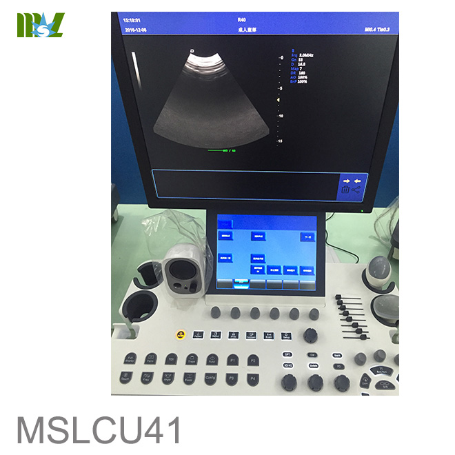
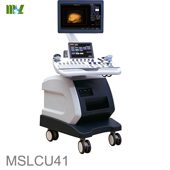
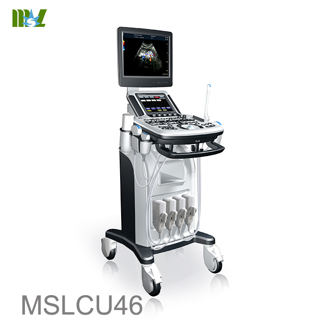
MSLCU41 High-End 4D color Doppler ultrasound system
|
MODEL
|
MSLCU41 3.0 VERSION
|
|
Computer specs
|
Windows Embedded operation system (CN,EN language)
19’’ medical monitor(1280*1024)+10.4’’ touch monitor
Intel i5 processor
4G RAM
120G SSD+500G HDD
|
|
Weight/Dimension
|
Unknown
|
|
Imaging Modes
|
2D, 3D, 4D, Color/PW/CW/Power/Directional Color Power Doppler,
Tissue Doppler, Color M-Mode, Free Steering
(Anatomical) M-Mode
|
|
Features
|
Compound Imaging,
Speckle Reduction Imaging,
Tissue Harmonics Imaging,
4D,
Automatic Image Optimization,
Tissue Doppler,
Image optimization, Multi-Beam, IMT, Trapezoidal imaging
iBank database
|
|
DICOM Modes
|
Store, Print, Working list, Storage Commitment, Structured Reports
|
|
Export Options
|
DICOM, Ethernet, JPG/BMP/PNG,AVI,
Network Storage, USB Memory Stick.
USB DVD/CD+R(W)
|
|
Input/Output
|
VGA, 2 USB Ports, Ethernet, Dicom, Built-in speakers
|
|
Transducer Types
|
Convex, Linear, Sector Phased, Micro Convex,
4D Volume Convex, Endocavity, Veterinary linear
|
|
Applications
|
Abdominal, OB/GYN, Urology, Cardiac, Vascular,
Small Parts, Pediatric, MSK
|
|
Probe Ports
|
4 Active
|
|
Cine Memory
|
>10 seconds, 750frames
|
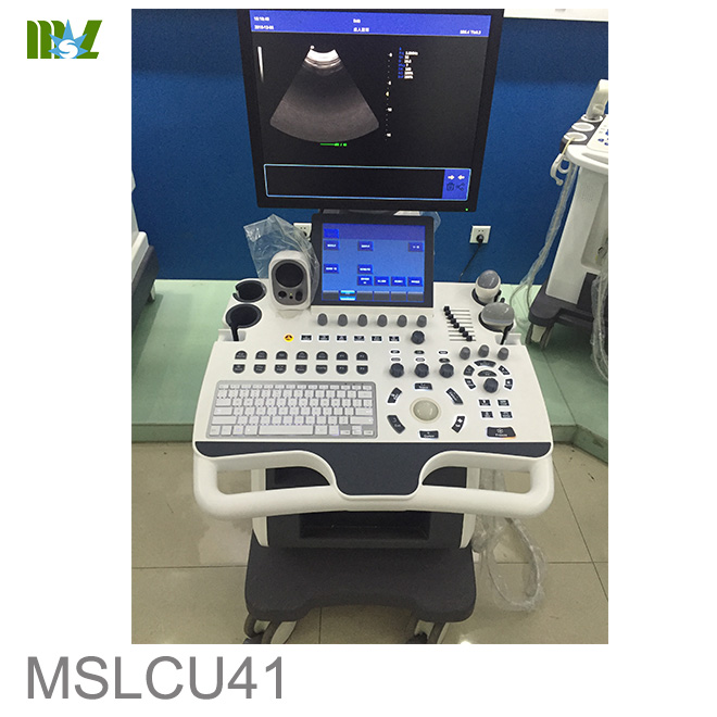
MSLCU41 High-End 4D color Doppler ultrasound system
1.Advanced Clinical operation:
i.Based on Windows XP operating system menu, with VGA 10.4 inch LED touch screen operating menu, English/Chinese lauguages for option, Screen layout can be edited, to increase or decrease menu quantity or change the display position as per users request, which is convenient for doctors to see current operation.
ii.One key automatic optimization (AUTO): Doctors just need to click AUTO key, the system will automatically adjust and optimize various imaging parameters according to the echo signal of different organization, such as this ultrasound system can be used to automatically optimize the spectrum and adjust the baseline and the pulse repetition frequency, instantly get satisfactory ultrasound images, make image adjustment more convenient and quick, improve the working efficiency of the clinical diagnosis.
iii.Parameter preset (organization characteristic imaging features) : doctor can make the best parameters of instrument themselves in the system set in advance according to imaging characteristics of different organizations, such as the liver, bravery, kidney, uterus, ovary and so on all sorts of viscera, in routine ultrasound diagnosis, to diagnose different organizations, doctors just need to choose preset key, the system will be fully automatic adjustment to the state of ultrasonic diagnosis for the organization, not need to adjust again, you will get an ideal image, which can improve the work efficiency.
iv.Custom shortcuts parameter setting function: five custom shortcuts buttons, users can define the use function of the five buttons, a key to realize intelligent operation, avoid the instrument parameter adjustment, improve the diagnostic speed.
v.Intelligence amplification function: can enlarge any ultrasonic area of interest, observed clearly magnified diagnosis, improve the working efficiency of the clinical diagnosis
vi.Color hiding technology: in color mode, do not need to exit the color mode can make the color hidden, allows doctors to quickly implement color image contrast observation and 2D structure
vii.RTSA real-time spectrum analysis function: D mode, (under scanning mode) can automatically envelope spectrum and calculate PSV, EDV, AVp, AVm, ats, DT, RI, PI and other hemodynamic parameters.
viii.Browse window function: the diagnosis of the current saved image, keep browsing the image on the left side of the screen, allows doctors to browse at any time, do comparison and analysis.
ix.Host interface can move around, with embedded keyboard and background light, can input Chinese/English information, realize the man-machine interactive
x.Task lamp navigation system: automatic analyze current activated function keys, allows doctors to clearly know current task, guide the doctor the next steps with multiple color indicator.
MSLCU41 High-End 4D color Doppler ultrasound system
2.Advanced Technology
i.A variety of high frequency probe configuration, the scope of probe frequency width: 2-14 MHZ, high-frequency linear array probe frequency is 14.0 MHZ, maximum satisfy the needs of clinical application.
ii.System dynamic range up to 260 db, including 15-145 db visible adjustable, in order to obtain clear and lay the foundation of 2D images.
iii.Using compound imaging technology, image resolution: longitudinal ≤1 mm, horizontal ≤ 0.5 mm,
the 2D image is exquisite, clear,easy to find early small lesions, help doctors to improve the diagnostic accuracy.
iv.Equipped with broaden the pulse imaging technology, improve the penetration and contrast of the images, probe scanning depth ≥ 360 mm, especially suitable for obese patients and patients who is not easy to get good images.
v.Combined with broadening sector imaging technology, abdominal probe: scanning Angle ≥ 105 degrees, intracavitary probe: scanning Angle≥160 degrees, higher probe scanning Angle can observe a broader conditions of the specific viscera, to show more image information, especially for checking big viscera.
vi.Linear array probe trapezoidal imaging and 2D beam deflection technology: viscera can be observed with a wider range of specific conditions and effectively avoid the front cover, show more clearly of viscera behind tissue of strong shot, allow doctors to scan imaging on special parts which is difficult to get.
vii.Tissue harmonic imaging (THI): system has filtering harmonic technology;Reverse phase pulse harmonic technology;Broaden the pulse harmonic technology, three kinds of harmonic technology to realize pure tissue harmonic imaging, to obtain high quality of tissue harmonic image, at the same time, the abdomen, high frequency, harmonic imaging probes have functions such as cavity, harmonic 2 period of adjustable frequency, harmonic model three kinds of options, maximum limit satisfy the needs of a variety of clinical
viii.With adaptive speckle suppression technology, make clearer boundary between tissue and administrative levels to enhance image, show the complete contour complex pathological changes, easy to distinguish the early pathological changes, helps doctors to improve diagnostic accuracy, adapted to the imaging difficult patients ultrasonic intervention and treatment, and other special purposes
ix.color doppler imaging system with a full digital color doppler blood flow imaging, directional energy doppler imaging features, of which the PW pulse wave doppler and the CW continuous wave doppler,≤1 mm/s, the PWD maximum blood flow velocity measurement ≧ 12300 mm/s, the CW maximum blood flow velocity measuring ≥33200 mm/s, and blood sample width and position scope: width 0.5-40 mm, it is a high-grade whole body practical colour Doppler ultrasound system.
x.Combined real-time dynamic density beam scanning (HDB) technology with high sensitivity (HSCFM) blood flow imaging technology, make the equipment with extremely high flow sensitivity, each blood flow information can be caught accurately
xi.With color blood flow deflection technology: to avoid insensitive phenomenon between the direction of blood flow and ultrasound beam vertical flow, blood flow sensitivity is high, with various kinds of deflection Angle options.
xii.Real-time three synchronous unit: 2D and color doppler, spectral doppler can be displayed at the same time, easy for doctors to analyze contrast.
MSLCU41 High-End 4D color Doppler ultrasound system
3.System Configuration
i.Display mode: B, B\B, M, B\M, 4B, B\C, B\C\D, anoramic imaging, broaden the perspective imaging, imaging, trapezoidal composite imaging (SCI) model to B, space, color blood flow chart pattern, color energy graph mode, the direction of energy graph mode, bipolar tissue harmonic imaging mode, PW pulsed wave doppler imaging, the CW continuous wave doppler imaging, 3 d / 4 d imaging mode
ii.Full digital 2D gray-scale image
iii.Color Doppler flow imaging
iv.Directional color Doppler energy imaging
v.PW pulsed wave doppler imaging
vi.CW continuous wave doppler imaging
vii.Space compound imaging
viii.Wide scene imaging (Option)
ix.High resolution compound imaging
x.THI, available for convex,linear, cardiac and trans-vaginal probes
xi.Adaptive speckle suppression technology
xii.Linear array probe trapezoidal imaging technology
xiii.Convex array probe broadening perspective imaging technology
xiv.Color doppler 2D mode automatically optimize adjustment technology
xv.Real time three synchronization
xvi.PIP intelligence picture imaging mode
xvii.Real-time 3D imaging mode: equipment equipped with built in 4d (3d) real-time imaging module software packages, users can use corresponding (real-time 3d) volume 4d probe, to get perfect function of 4D imaging.
xviii.multi package, in addition to the general measurement software package, with abundant peripheral vascular, gynecology and obstetrics, heart, urologist, newborn, orthopaedic surgery, and so on measurement and analysis of special packages blood flow, maximum limit satisfy the needs of clinical.
xix.built-in E - COM graphic management system: 560GB hard disk storage, editable ultrasound diagnosis report in Chinese/English, embedded ultrasonic diagnostic image in the report, and directly storage, printing, callback, query and so on, burning built-in DVD driver and USB interface
xx.display: 19 inches LED high resolution display
xxi.multi probe options:
1)Convex probe: 2.5-5.0MHz (Variable frequency, harmonic frequency ≥5 kinds), Probe scanning Angle of 20 ° ~ 85 °, visual and adjustable.
2)Linear probe: 6.0-14.0MHz (Variable frequency, harmonic frequency ≥4 kinds), Probe scanning with trapezoidal imaging and 2D beam deflection technology.
3)Trans-vaginal probe: 5.0-9.0MHz (Variable frequency, harmonic frequency ≥2 kinds), Probe scanning Angle of 20 ° ~ 160 ° visual and adjustable.
4)With real-time 3d (4d) volume probe: 2.0-5.5 MHz, 4 kinds of frequency adjustable.
Optional phased array probe for cardiac: 2.0-5.5MHz, 5 kinds of frequency adjustable.
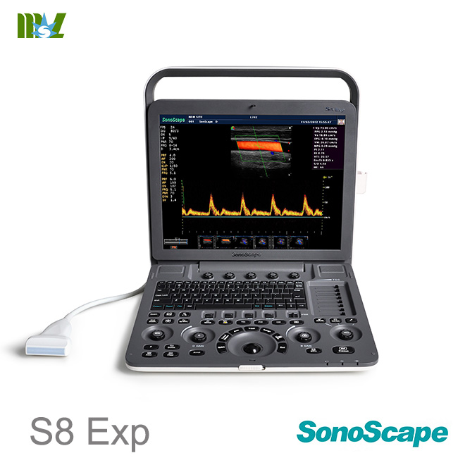
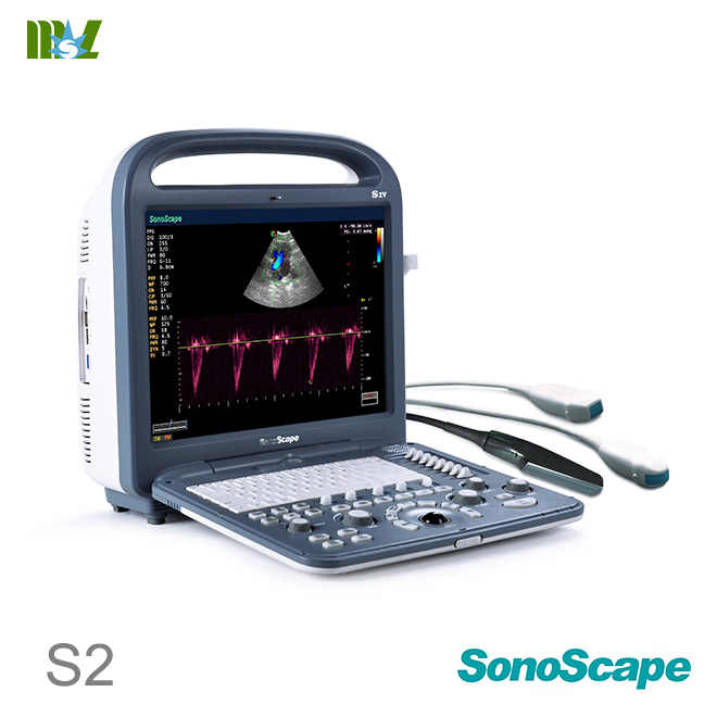
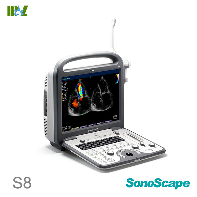
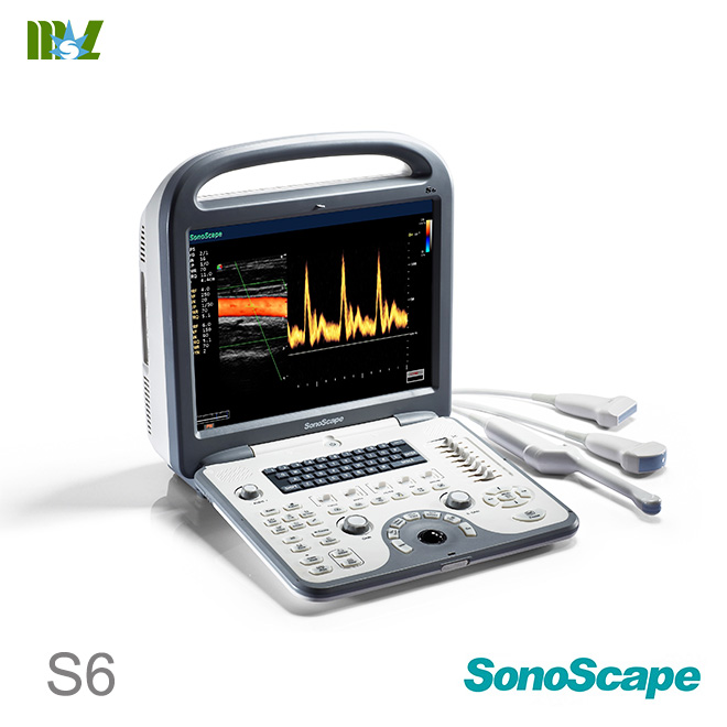
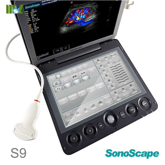
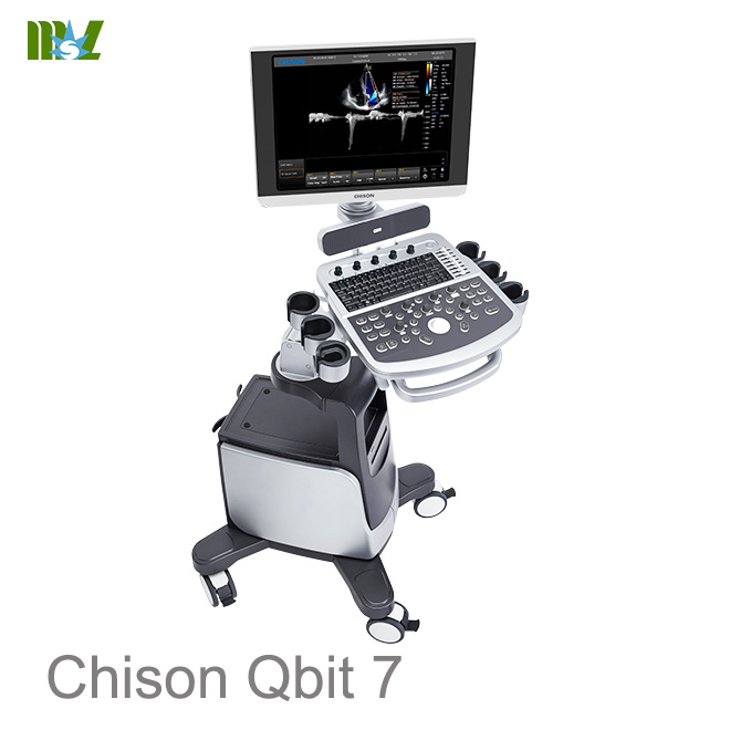
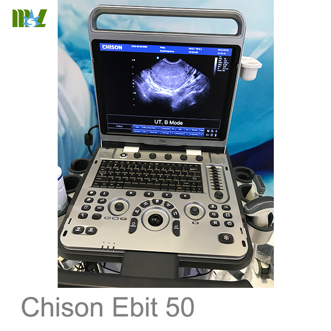
Hot sale Sonoscape ultrasound | Chison ultrasound price list
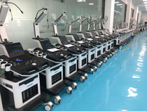
MSL TEAM picture


MSL Certificate
MSL Medical cooperate with DHL,FEDEX,UPS,EMS,TNT,etc.International shipping company,make your goods arrive destination safely and quickly.


 Price is 8-20% Lower Than Other