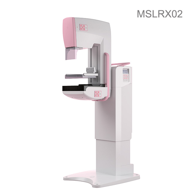
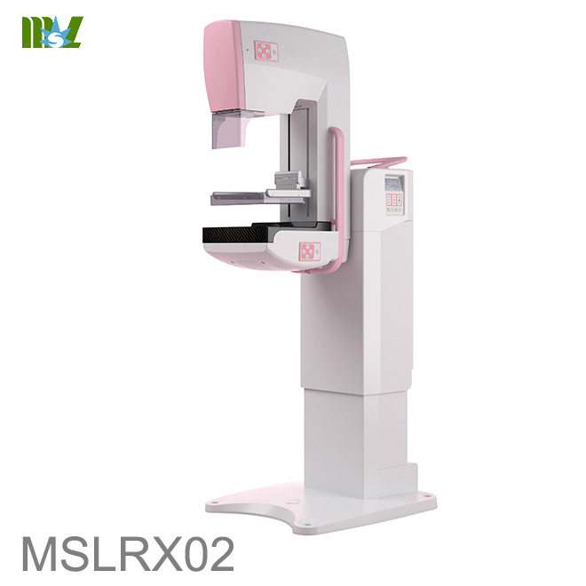
Efficient Navigator DR Mammography MSLRX02
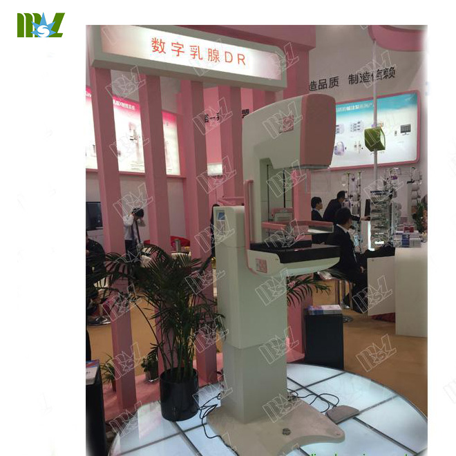
Perfect environmental adaption makes it available for indoor and vehicle application for different intended use.
User-friendly GUI(Graphic user interface),support various exposure mode
User-friendly Interface, simple and easy to operate
Fast transmission speed, comply with DICOM3.0 protocol, can achieve the seamless link with HIS. RIS. and PACS systems.
Powerful tools for patient management, data gathering and image reconstruction, viewing and measuring, typesetting, printing, storage, provides rich aides for diagnosis
With special detector of a-Si for breast mammography, X-ray is transferred to electric signals directly without the additional process of transferring x-Ray to visible and then to electric signal.This eliminates the artifact due to scattering of X-Ray and guarantees the authenticity from the imaging mechanism and can really satisfy the requirements of breast mammography on details. With high DOE, this can guarantee high resolution of image and meanwhile decrease irradiation dose.
Efficient Navigator DR Mammography MSLRX02
|
Item
|
Description
|
|
1
|
Mammography System
|
|
|
Rating power:5000VA
Integrated System (Generator Integrated within the main device)
High voltage generator output: ≥100 kHz, 20 kV to 40 kV increment step: 1 kV
High performance X-ray tube:
Standard:Molybdenum Anode, Focal Spot Size: 0.1 mm (Small)/ 0.3 mm (Large)
Optional:Tungsten Anode, Focal Spot Size: 0.1 mm (Small)/ 0.3 mm (Large)
Support isocenter rotation function
C-arm Rotation range: +180°~ -135°
Height range of breast support surface: 735mm-1315mm;
C-arm power-driven electrically, Dual side digital display;
Compression plate: flexible and multi-level smart Compression
Spatial Resolution: >6 Lp/mm
Detector type: Amorphous Silicon Detector
Effective Imaging Area: 24*30cm
Acquisition Station compatible with DICOM 3.0
Focal distance : 650mm
Added filter: Mo / Rh
Added filter Switch: Manual/Automatic
X-ray Field adjustment: Manual
Magnification factor: 1.5
Exposure mode: Manual / AEC
Supply: 220V~,±10%
Frequency: 50/60Hz±1Hz
Max. power : 5000 VA
N.W.: 240 Kg
Compact design, minimum device height: < 1.1 m
Packing Size: 121 X 94 X 145 CM
|
|
2
|
High-Voltage Generator
|
|
|
a) Frequency:≥100KHz;
b) Tube voltage Range: 20 kV ~ 40 kV in 1 kV step;
c) Max Output: 4 kW;
d) Max tube voltage:40kV;
e) Max tube current: 140mA;
f) Max current time product: ≥630mAs;
g) Ripple: <4%.
|
|
3
|
X-Ray Tube
|
|
|
Standard:
Imported X-ray tube for Mammography;
Anode Type: Molybdenum;
Focal Spot Size: 0.1 mm (Small)/ 0.3 mm (Large)
Permanent filtration: 0.5mmBe
Max anode speed: 10000rpm/min;
Max tube voltage:40kV;
Max tube current: 35mA(small)/140mA(Large)
Anode heating capacity: 300 kHu
|
|
Optional:
Imported X-ray tube for Mammography;
Anode Type: Tungsten;
Focal Spot Size: 0.1 mm (Small)/ 0.3 mm (Large)
Permanent filtration:0.5mmBe;
Max anode speed: 10000rpm/min;
Max tube voltage:49kV;
Max tube current: 35mA(small)/140mA(Large)
Anode heating capacity: 300 kHu
|
|
4
|
Beam Limiting Device
|
|
|
Electric switch between Molybdenum or Tungsten added filter;
Adjustable X-ray field manually.
Light field indicator is on automatically as compression plate moves down;
Light field indicator is off with delay as compression plate stops moving;
e) Off delay of light field indicator can be set via software.
|
|
5
|
C-arm Assembly
|
|
|
C-arm up-down and rotation movement controlled via C-arm control panel;
C-arm stops automatically at common mammographic position: CC, OBL and LAT;
Rotation angle of OBL position can be set via software;
Support table up-down range: 735mm(L)~1315mm(H);
Rotation type of C-arm: isocenter rotation around the examined part;
Rotation range of C-arm: -135o~180o
Distance between focal spot and image reception area:650mm;
Magnification factor:1.5
Device can be installed in the movable physical vehicle as max height of device is less than 110cm;
Local display panel on the both side of the stand can indicate rotation angle of C-arm;
For safer operation, emergency stop button is locate on the top of C-arm;
Duplex foot switch(2 pcs) provided.
|
|
6
|
Compression Device
|
|
|
Up-down movement : power-driven electrically;
Compression control: continuous activation by foot;
Compression mode: flexible and multi-level compression, automatic and manual decompression;
Compression plate travel range: 5~268mm;
Compression force: 0~200N
Compression thickness: 5~268mm
Size of compression plate: 24X30cm.
|
|
7
|
Local Operation and Display
|
|
|
a) Dual display with backlight on the both side;
b) Parameters displayed:
- rotation angle of C-arm;
- compression thickness;
- compression force;
- KV;
- mAs;
- density
- focal spot selected;
- exposure mode;
- Added filter selected;
- automatic decompression indication;
- state indication;
- failure alarming indication;
c) Control function:
- KV;
- mAs;
- density
- selection of focal spot;
- selection of filter;
- selection of mammographic mode;
- selection of automatic decompression after exposure.
|
|
8
|
Anti-scatter Grid
|
|
|
Support movable grid;
Grid ratio: 5:1;
Grid density: 41 L/cm;
Grid focal distance:65cm;
Manually unload grid in magnification mode.
|
|
9
|
Digital Image Detector
|
|
|
Detector type: Amorphous Silicon Detector;
Material: Amorphous Silicon + CSI;
Min. pixel: 85µm;
Image Matrix Sizes: 2816×3528 pixels;
Effective Imaging Area: 24cm×30cm;
Image Reading Duration: <1.4s;
Interval Duration: <30s;
Spatial Resolution: 6 lp/mm;
MTF: 50%;
DQE: 65% (1 Lp/mm);
Output Gray scale: 14 bits
|
|
10
|
Acquisition Station
|
|
|
Intel CPU frequency≥2.4GHz,≥ 4 GB Memory, ≥500 GB HDD
LCD Display
Image Acquisition Software
Image Detector Self-check and adjustment
Patient Information and Image Management
Auto Explore Control
Acquisition of Explored Image Information
Browsing and Searching of Image
Measure the Distance, Angle, Space of Image and Mark The Selected Area
Image Process after Acquisition: Black and white reverse, rotate and flip, zoom, local editing, enhancement, filtering noise, copies, abridged, etc.
DICOM 3.0 Services (Print, Store, Query/Retrieve, CDRW, Scheduled Workflow and Patient Information Reconciliation)
Automatic failure diagnosis, error code and message indication;
Exposure activation: exposure button;
Data backup and recovery.
|
|
11
|
Automatic Exposure Control
|
|
|
Automatically select imaging parameters according to the thickness of breast and density of tissue;
Integration detector;
Automatically position selection.
d) Automatically kV, filter and mAs selection.
|
|
12
|
Manuals
|
|
|
User Manual
Pre-Installation Manual
|
|
Item
|
Description
|
|
1
|
High resolution medical image display
|
|
|
Display Hardware: 5MP LCD Display at 2K x 2.5K
|
Efficient Navigator DR Mammography MSLRX02
|
Power Capacity
|
5000VA
|
|
Voltage
|
220V~,±10%
|
|
Frequency
|
50/60Hz±1Hz
|
|
Earth resistance
|
≤0.6Ω
|
|
Item
|
Temperature
|
Humidity
|
Atmosphere Pressure
|
|
Shield Room
|
15℃~35℃
|
30%~60%
(No dewing)
|
700hPa~1060hPa
|
|
Operator Room
|
10℃~40℃
|
20%~80%
(No dewing)
|
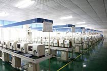
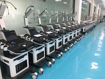
MSL TEAM picture


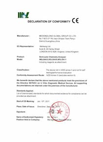
MSL Certificate
MSL Medical cooperate with DHL,FEDEX,UPS,EMS,TNT,etc.International shipping company,make your goods arrive destination safely and quickly.


 Price is 8-20% Lower Than Other