


 Price is 8-20% Lower Than Other


What Is USG?
USG is Diagnostic sonography (ultrasonography) is an ultrasound-based diagnostic imaging technique used for visualizing internal body structures including tendons,muscles,joints,vessels and internal organs for possible pathology or lesions.The practice of examining pregnant women using ultrasound is called obstetric sonography,and is widely used.
How Does USG Work?
Today Medsinglong let you know what is Medical ultrasonography(USG).
In physics,'ultrasound' refers to sound waves with a frequency too high for humans to hear.Ultrasound images (sonograms) are made by sending a pulse of ultrasound into tissue using an ultrasound transducer (probe).The sound reflects (echoes) from parts of the tissue; these echoes are recorded and displayed as an image to the operator.
Many different types of images can be formed using ultrasound.The most well-known type is a B-mode image,which displays a two-dimensional cross-section of the tissue being imaged.Other types of image can display blood flow,motion of tissue over time,the location of blood,the presence of specific molecules,the stiffness of tissue,or the anatomy of a three-dimensional region.
Compared to other prominent methods of medical imaging,ultrasonography has several advantages.It provides images in real-time (rather than after an acquisition or processing delay),it is portable and can be brought to a sick patient's bedside,it is substantially lower in cost,and it does not use harmful ionizing radiation.Drawbacks of ultrasonography include various limits on its field of view including difficulty imaging structures behind bone and air,and its relative dependence on a skilled operator.
The ultrasound image is produced by sending sound waves of 1-10 million hertz through a transducer by placing it over structures of the body. The sound waves are either absorbed or bounce back to crystals in the head of the transducer. For example, sound waves go through areas that are hollow or fluid-filled, such as the bladder and blood vessels. These areas appear black on the screen. Areas filled with tissue allow some penetration and refraction of sound and produce a grayish-white image. Really hard structures,such as bone, produce a bright white image as the sound waves completely bounce back to the transducer.
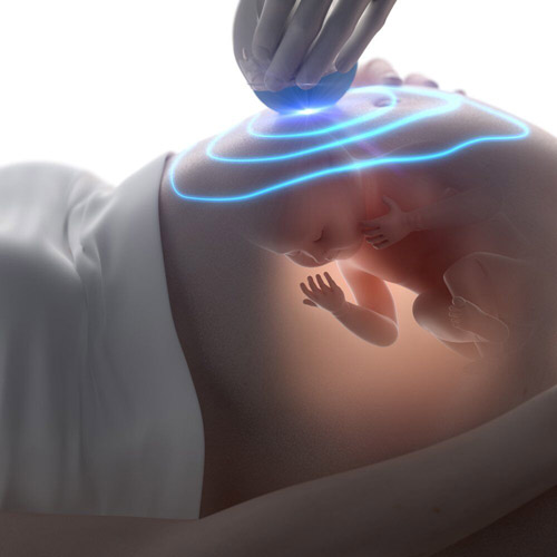
In pregnancy, this allows providers to have an image of the woman's womb. The amniotic fluid will appear black, which enhances the bones and tissues of the baby, which will appear white. The doctor explains to Patty that the ultrasound is ordered so he can assess how well the baby is developing, determine the gender of the baby, and detect any abnormalities. He can also measure Patty's cervix and ovaries.
USG Technology:
1.Universal Charging System,or Universal Charging Solution,an agreement on requirements for a common External Power Supply within Open Mobile Terminal Platform.
2.Medical ultrasonography,an ultrasound-based diagnostic imaging technique used to visualize internal organs.
3.Urine specific gravity (kidney),a measure of urine concentration or relative density
History of Ultrasonography
We can thank the French for the invention of ultrasound. Physicist Pierre Curie first used ultrasound in 1877. Imaging was discovered by Paul Langevin 35 years later. Ultrasound was first used during wartime to detect submarines and wreckage in the ocean. It was used medically in the 1920s for physical therapy for Europe's soccer teams. Austrian physician Karl Dussik and his brother Friedrich Dussik were the first to use ultrasound for diagnostic purposes, such as looking at brain structures, in the 1940s.
Real-time ultrasound, similar to the one Patty will have, wasn't developed until the 1980s. This is when actual images could be seen and interpreted. The 90s led to 3D and 4D imaging where the public was able to be viewed and it was possible to recognize familiar images such as facial features of a baby.


