


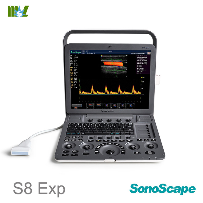
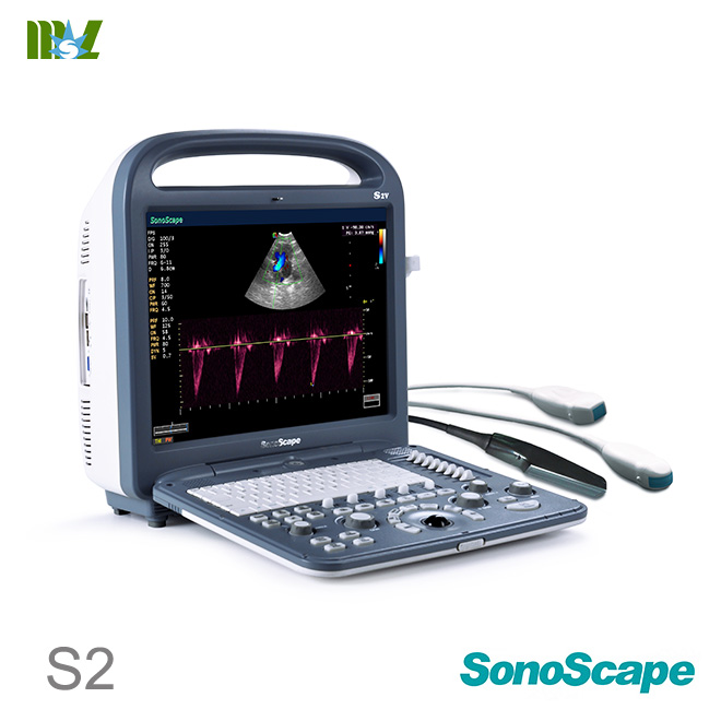
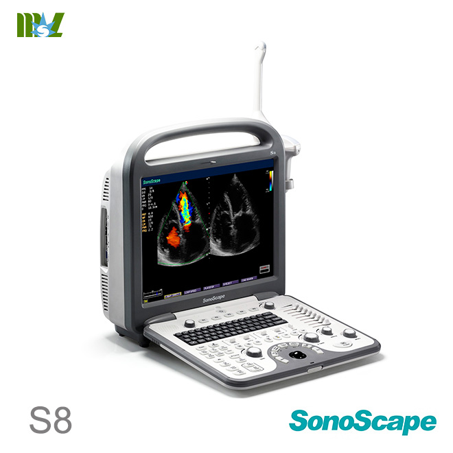
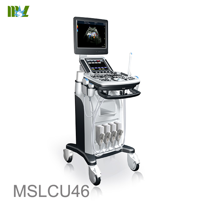
 Price is 8-20% Lower Than Other
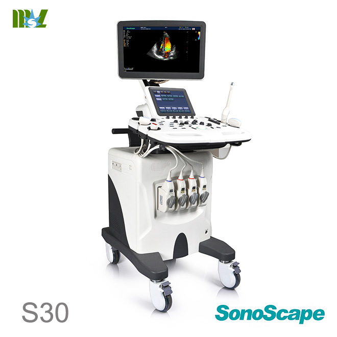


M-mode ultrasound (also called motion-mode imaging) does not yield full frame images per se, but rather one selected image line is rendered as a function of time. This is used for displaying motion of, for example, the periodic movement of heart valves. Any abnormalities or temporal variations can be directly seen as an image on the screen. The B-mode cross-section of a carotid artery is shown in Fig. 13a. Proximal and distal vessel wall delineates the dark vessel interior, as indicated by the arrows to the right.
In a M-mode representation in Fig. 13b, pixels along the white vertical line in (a) are repeated parallel to each other over time. Figure 13b shows 5 s of repeated scans. For each heart beat a pulsatile wave travels through the arterial blood pool locally expanding the blood vessels. This expansion can be seen in B-mode as well as in M-mode representation.
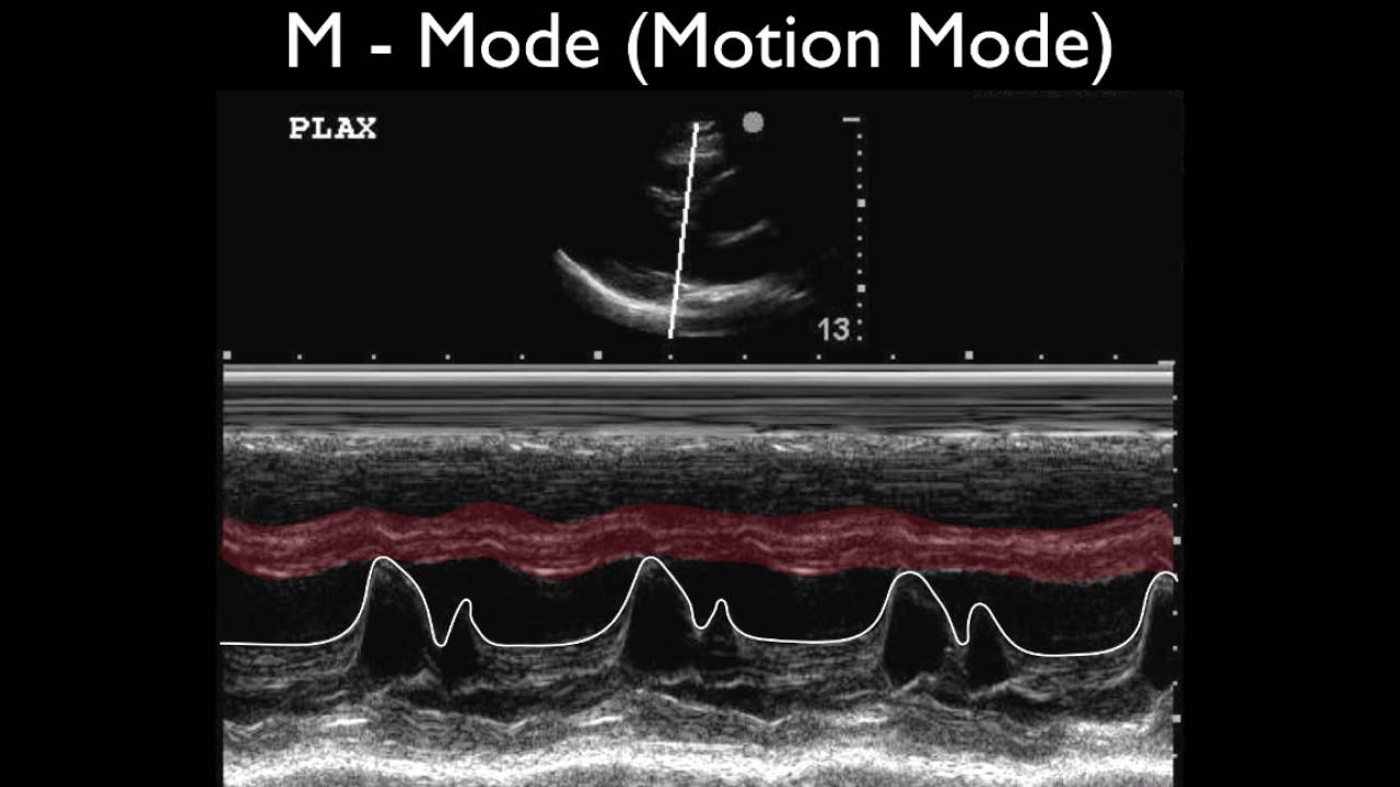
However, in B-mode it is an event in time occurring over several image frames, whereas in M-mode this event is plotted as the horizontal axis and therefore easy to detect. White arrows in Fig. 13b indicate the temporal expansion of the blood vessel. Figure 13c shows a much more pronounced motion. The transducer was pointed toward the heart and is therefore either imaging the heart wall or one of the heart valves, showing the typical cardiac pattern.


MSL TEAM picture


MSL Certificate

MSL Medical cooperate with DHL,FEDEX,UPS,EMS,TNT,etc.International shipping company,make your goods arrive destination safely and quickly.

Welcome to medicalequipment-msl.com,If you have any demand in ultrasound machine.
Please feel free to contact cindy@medicalequipment-msl.com.


