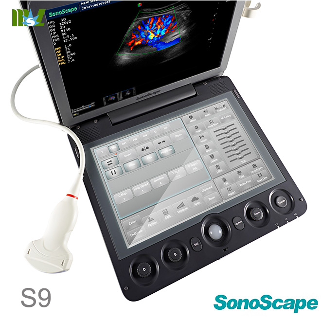
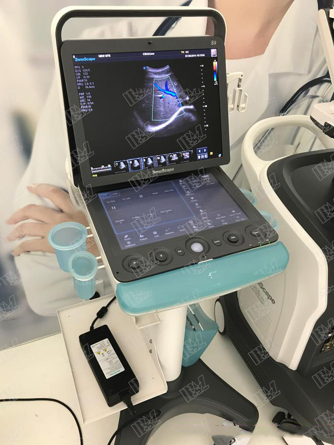
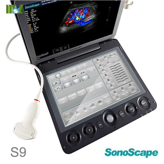
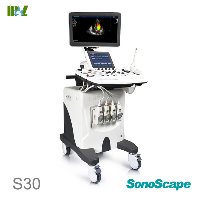
The S9 Color Doppler Ultrasound System adopts the advanced ultrasonic Doppler technologies, including the Full Digital Super-wide Band Beam Former, Digital Dynamic Focusing, Variable Aperture and Dynamic Tracing, Wide Band Dynamic Range, Multi-Beam Parallel Processing, etc. The ultrasound diagnostic software system, ultrasound system imaging, multi-languages operation interfaces and touch screen with human-computer interaction technology can be customized easily in accordance with the design of human engineering. Users can perform the system with the minimum requirement of training or guidance. This system has been designed to comply with applicable international standards and regulations, ensuring the safety and availability of this product. This system is based on the computer technology and Linux operation system, which make the system more flexible and stable. System maintenance and function update can be completed by software updating, through which would promote product value and keep the technological advancement.
ecografie transfontanelara SonoScape S9 Advanced Technologies
* New generation digital front-end
technology
* Multi-beam processing technology
* Spatial compound imaging
* Post-processing technology
* Tissue Harmonic Imaging
* High Pulse Repetition Frequency
* Panoramic imaging
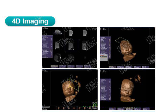
* 4D imaging
* Graphic diagnosis icon
* Touch screen with human-computer
interaction technology
ecografie transfontanelara SonoScape S9 Standard Configurations
* B mode
* Color mode
* PW mode
* CW mode
* THI mode
* DPI mode
* DDPI mode
* 3D imaging
* Cardiology measurement package
* Gynecology measurement package
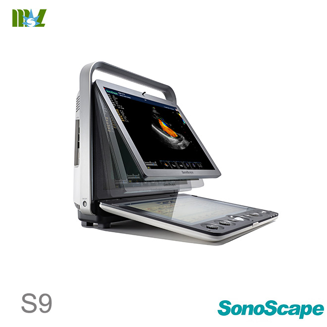
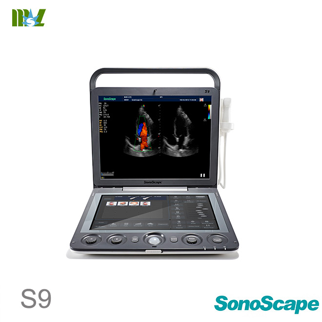
* Urology measurement package
* Vascular measurement package
* Small parts measurement package
* Orthopedic measurement package
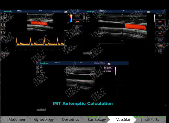
* IMT measurement
* TEI index
* Spectral Doppler auto trace
* Color flow volume measurement
* MLA probe
* Phased array probe
* Multi-beam processing technology
* μ-scan function
Specifications for S9 Color Doppler Ultrasound System
2
* TDI function
* Steer M mode
* Color M mode
* B mode: Five variable frequencies
* High Pulse Repetition Frequency
* Triplex mode
* Panoramic imaging
* Compound imaging
* Trapezoid imaging
* ECG function module
* DICOM transmission
* DICOM storage commitment
* DICOM worklist function
* DICOM MPPS
ecografie transfontanelara SonoScape S9 Optional Functions
* 4D imaging
* TEE probe
* Image rotation function
* B Flow function
* Stress echo
ecografie transfontanelara SonoScape S9 Optional Accessories
* Biopsy guide
* Color ink-jet printer
* B/W video printer
* Color video printer
* Probe cable hanger
* Foot switch
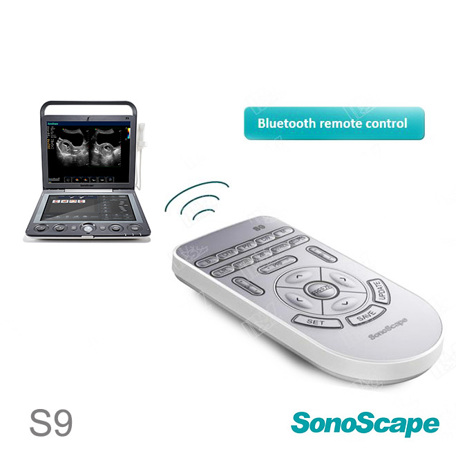
* Remote control
* Trolley
ecografie transfontanelara SonoScape S9 Probe Scan Ranges
* Curved transducer: ≥70°
* Phased array transducer: ≥90°
* Micro-curved transducer: ≥193°
ecografie transfontanelara SonoScape S9 Scan Methods
* Electronic curved sector scan
* Electronic linear array scan
* Electronic phased array sector scan
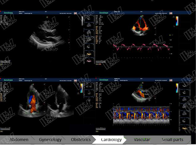
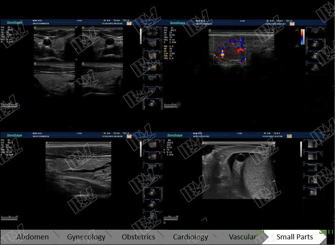
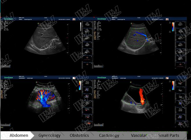
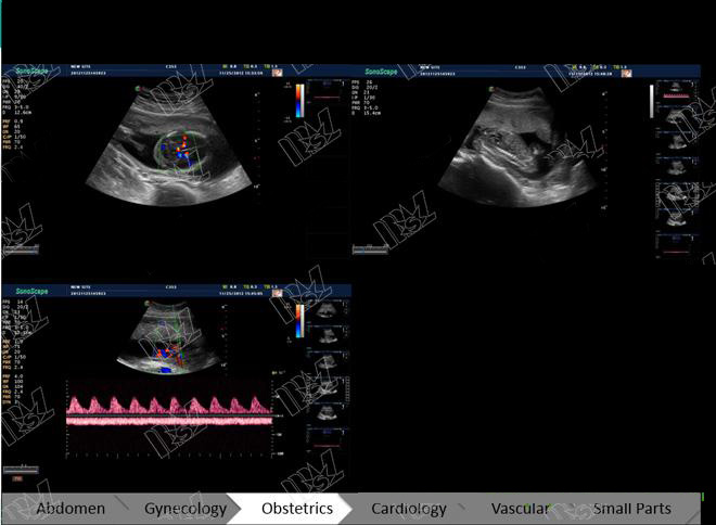
ecografie transfontanelara SonoScape S9 Applications
* Abdomen
* Vascular
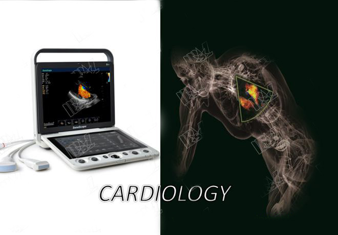
* Cardiology
* Gynecological/Obstetrical
* Urology
* Musculoskeletal
* Interventional ultrasound
* Small parts
* Anesthesiology
* Pediatrics
* Emergency
ecografie transfontanelara SonoScape S9 Imaging Modes
* B mode
* M mode
* THI mode
* CDI mode
* DPI mode
* TDI mode
Specifications for S40 Color Doppler Ultrasound System
3
* PW mode
* CW mode
* 3D/4D mode
* Color M mode
* Steer M mode
ecografie transfontanelara SonoScape S9 Display Formats
* Dual B
* Quad B
* B + PW
* B + CW
* B + M
* B + Color
* Dual B (Flow)
* B + Color + PW
* B + Color + CW
* B + Color M
* Panoramic imaging
* Trapezoidal imaging
ecografie transfontanelara SonoScape S9 System Configuration
Menu
* Exam History
* New Exam
* Continue
* Review
* Select All
* Store to DICOM
* Store to USB
* Delete
* Print
* Report
* Exit
* Delete
* Store
* Print
* Commit
* Exit
* System Settings
* General Settings
* Facility Name
* Language
* English
* Simplified Chinese
* Spanish
* Russian
* French
* Italian
* German
* Turkish
* Screen Saver
* Trackball Sensitive
* Clip Format
* MP4, AVI
* Still Format
* JPG, BMP, TIF
* Screen Save: adjustable
* Color of ROI
* Green, Yellow
* Orange, Blue
* Display Format:H1/2, H1/4, V1/3,
V1/2, V2/3, O1/4
ecografie transfontanelara SonoScape S9 VIDEO
Specifications for S9 Color Doppler Ultrasound System
4
* One Key Save: On/Off
* EFW Unit: selectable
* Date Format
* mm /dd/yyyy
* yyyy/mm/dd
* dd/mm/yyyy
* Report Format
* PDF, TXT
* Save Frame Number: adjustable
* Set Printer
* Printer Driver
* Video Invert
* Insert Driver
* Set Calculation Menu
* 2D Mode
* Angle
* Volume
* Volume L×W×H
* Doppler Area
* Color Flow
* IMT
* Vascular
* Small Part
* Orthopedic
* Obstetrical/Gynecological
* Left Ventricle
* Urologic
* Carotid artery health evaluation
* Carotid artery health evaluation
(CF)
* Mitral Valve Diameter
* Lv Outflow Diameter
* Pul.Valve Diameter
* Aorta Diameter
* PW Mode
* Flow Velocity
* Acceleration
* Time
* Heart Rate
* Cardiac
* Obstetrical/Gynecological
* Vascular
* Carotid artery health evaluation
(PW)
* M Mode
* Distance
* Time
* Slope
* Heart Rate
* Left Ventricle
* Mitral Valve
* Aortic Valve
* Set Measurement Method
* BSA setting
* Eastern
* Western
* Measure Method
* Ellipse
* Trace
* Package
* All Packages
* Continue Dist: On/Off
* Dop Auto
* AUTO
Specifications for S40 Color Doppler Ultrasound System
5
* SEMI-AUTO
* Focal Auto: On/Off
* EFW Method
* WEI/SAB HC, AC, FL
* Shepard AC, BPD
* Hadlock1 AC, FL
* Hansman AC, FL, HC
* Tokyo BPD, APTD, TTD, FL
* Hadlock2 HC, AC, FL
* Hadlock3 BPD, AC, FL
* Hadlock4 HC, AC
* Hadlock5 BPD, HC, AC, FL
* Shinozuka BPD, AC, FL
* Warsof FL,AC
* Campbell AC
* Mediscan FL, AC
* Mediscan BPD, AC
* BPD Method
* Hadlock
* Jeanty
* Crespigeny
* Kurtz
* Hansmann
* Sabbagha
* Campbell
* Tokyo
* Merz
* Osaka
* FL Method
* Hadlock
* Hohler
* Jeanty
* Hansmann
* Tokyo
* Merz
* Chitty
* Osaka
* Campbell
* CRL Method
* Robinson
* Hadlock
* Nelson
* Jeanty
* Hansmann
* Mediscan
* Tokyo
* Osaka
* AC Method
* Hadlock
* Hansmann
* Tokyo
* Merz
* Campbell
* TAD Method
* Hansmann
* OFD Method
* Hansmann
* HC Method
* Hadlock
* Jeanty
* Chitty (M)
* Chitty (D)
* Merz
* Campbell
Specifications for S9 Color Doppler Ultrasound System
6
* GS Method
* Nyberg
* Hansmann
* Hellman
* Tokyo
* China
* Fibula Method
* Merz
* Radius Method
* Merz
* Mediscan
* Humerus Method
* Jeanty
* Merz
* Osaka
* Ulna Method
* Jeanty
* Merz
* Mediscan
* Tibia Method
* Jeanty
* Merz
* AUA Result by
* Average
* Last
* User define OB Method
* Replace
* Save
* Cancel
* Annotation Edit
* Insert
* Delete
* Edit
* Save
* Define Quick Key (0-9)
* OB measurement
* Cardiac measurement
* Load Default
* Load
* Create
* Retrieve
* Copy user setting to USB
* Copy user preset to USB
* Load USB user setting to system
* Load USB user preset to system
* DICOM Settings
* Local
* Store
* Worklist
* Print
* MPPS
* Commit
* System Info
* Control Number
* Software Version
ecografie transfontanelara SonoScape S9 System Parameters
* Frame rate: max. 750fps
* Grayscale Level: 256
* Transducer Elements: Max. 256
Specifications for S40 Color Doppler Ultrasound System
7
13. B Mode
* Gain: 1-255 adjustable
* Scan Depth: 32.9cm
* Image Zoom, Showing zoom ratio
* TGC: 8 levels slider controls
* Image Inversion: Left and Right, Up and
Down
* Panoramic imaging: achievable
* Compound imaging: adjustable
* Focus: Up to 12, Focus span adjustable
* Frequency: 5 bands adjustable
* Chroma: 13 types selectable
* Adaptive image fusion: 15 types
selectable
* μ-Scan: adjustable
* Line Density: 3 levels adjustable
(High/Medium/Low)
* Fame Relativity: 0-95 selectable
* Biopsy Guide Function: On/Off
* Biopsy lines angle adjustable
* Biopsy lines offset adjustable
* Dynamic Range: 20-280 (Probe
dependent)
* Grayscale Curve: 7 selectable
* Imaging width and position: adjustable
* Power: 1-100 adjustable, one step each
* TDI Type:1400-1700
* Trapezoid Imaging: On/Off (Linear array
probe)
* B steer Mode (Linear array probe)
14. Color/TDI Doppler
* Gain: 0-255
* Frame Rate: ≥50 frames/sec
* Size and position of color ROI: adjustable
* Auto Focus (focus number:1)
* Inversion: Up/Down, Left/Right
* Flow Invert: On/Off
* Frequency Range: 5 steps, adjustable
* Wall Filter: 25-750Hz, adjustable
* PRF: 0.5-12kHz (Probe dependent)
* Line Density: 4 kinds
(low/medium/high/super-high)
* Color/Direction energy: 11 kinds,
selectable
* Color baseline adjustment: ±15 steps
* Persistence: 0-80 (Probe dependent)
* B Reject: 0-255
* Linear deflection angel: 0, ±16, ±20
adjustable
* Flow profile: Achievable in Freeze Mode.
15. M Mode
* Steer M: 3 sample lines, Display frame
rate
* Video Inversion (On/Off)
* Chroma: 5 types
* Display Format: H1/2, H1/4, V1/3, V1/2,
V2/3, O1/4
* Scan Speed: 6 levels adjustable
* M Processing: Switch between average
and peak values
* Power: 30-100 adjustable
Specifications for S9 Color Doppler Ultrasound System
8
16. Spectral Doppler
* Doppler Methods
* PW (pulsed wave) Doppler
* CW (continuous wave) Doppler
* 2D Refresh: On/Off
* Sample volume and position for PW
Doppler: 1-20mm adjustable
* Video Inversion: On/Off
* Spectrum Inversion: Achievable
* θ Angle Correction: On/Off (correction
range: 0-80°)
* Spectral Real-time Trace: Achievable
* Baseline Shift:17 steps selectable
* Frequency Range: 5 steps
* Wall Filter: 25-750 adjustable
* PRF: 1~20kHz (PW)
* PRF: 1-48KHz (CW)
* Max Velocity Range:
* 0.0004-40.9 m/s (PW)
* 0.0013-49.1 m/s (CW)
* Scan Speed: 2, 4, 6, 8 sec/plane
* Doppler Chroma: 5 kinds selectable
* One-key Auto Optimization
* Auto Adjusting Baseline
* Auto Adjusting PRF
* Auto Correcting Angle
* Dynamic Range: 10 types selectable
* Display Format:H1/2, H1/4, V1/3, V1/2,
V2/3, O1/4
* Deflection Angle: 0, ±16, ±20,5 levels
adjustable
17. 3D/4D Mode
* Display Mode:
* Double Planes
* Quad Planes
* 3D Full Display
* 4D Full Display
* Crop Plane: On/Off
* Undo Cut
* X Rotation
* Y Rotation
* Z Rotation
* Horizontal Movement: Left/Right
* Vertical Movement: Up/Down
* Zoom Function
* Trace Cut: On/Off
* Render Mode: Vol, MaxIP, X-ray
* Auto Rotate: 45°, 90°, 180° and 360°,
selectable
* Opacity Offset: 0-255 adjustable
* Opacity Slope: 0-255 adjustable
* Scan Mode: Lin and Sec, selectable
* Zoom In/Out: adjustable
* Z Axis Angle: 10°-170° adjustable
* Color Map: 4 types
* Slice: A, B and C planes
* Slice Spacing: 0.5-2.0, adjustable
* Rescan: On/Off
* Stability: adjustable
* Scan Angle: 20°-75° adjustable
* Image Quality: High, Medium and Low,
adjustable
Specifications for S40 Color Doppler Ultrasound System
9
* 4D Image Gain: adjustable
* Frame Rate: 5f/s
* Cutting line curvature and position:
adjustable
* Copy frame size and position: adjustable
* Volume Playback: 0-9 adjustable
* 3D Image Storage
* 4D Image Storage and playback
* Print Function
18. Physiological Signal
Display
* ECG Pulse wave
* ECG Lead-three lead system
* ECG Gain: adjustable
* ECG Position: adjustable
* ECG Invert: On/Off
* R-Trigger: On/Off
* Trigger Delay: adjustable
* Frame Count: adjustable
19. Integrated Data
Management System
* Digital Channel: 1024
* Hard Disk Memory Capacity: More than
320G
* USB Interface: 3 (including 1 engineering
interface)
20. Image Storage and
Playback
* Cine loop: Up to 500 frames in B mode
* Cine loop time: 50 seconds or more
* Real time single/dual static and dynamic
image storage
* The stored images can be viewed directly
on PC.
* Crop board function: Achievable in freeze
state of B mode
* Doppler cine playback: Speed is
adjustable; Sound can be played back.
21. DICOM Network
Communication
* Storage: Directly transmits images with
patient information to a DICOM file server
* Print: Images can be printed directly
using a DICOM compatible printer
* Medical digital images and
communication DICOM 3.0 interface
22. Preset Function
Users can customize the presets based on
different probe and diagnostic part to optimize
imaging parameters and adjustment
combination.
23. Patient Data Management
* Patient Registration: Name, ID, Gender,
Date of Birth, Height, Weight, LMP, EDD
and GA.
Specifications for S9 Color Doppler Ultrasound System
10
* Patient Data, Report, Images can be
searched, played back and printed
24. Annotation and Body Mark
Setting
* Body Mark Icon: More than 52 kinds
* Annotation can be selected in the library.
* Annotation Number: Up to 20
25. Size
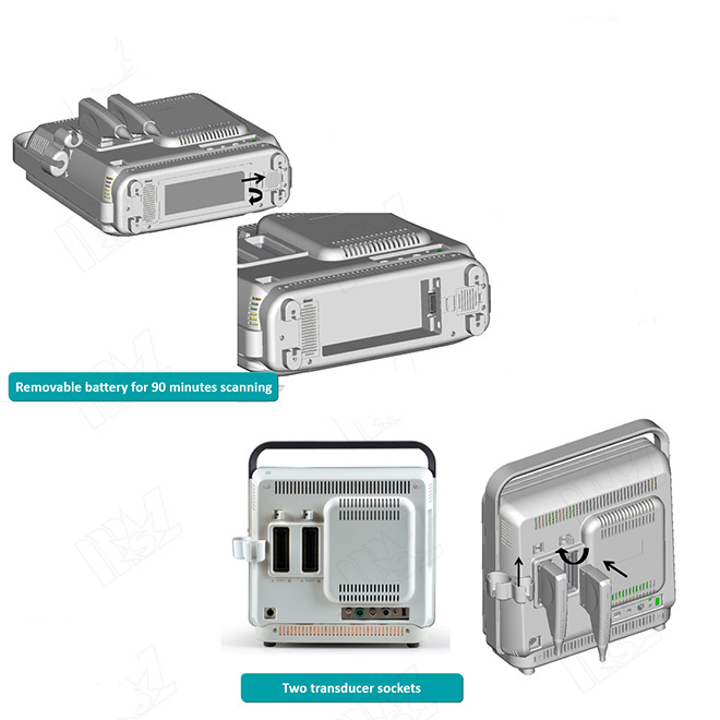
* L×W×H(mm): 137×357×392
26. Weight
* Weight: Approx. 7.8Kg
27. Probe Connectors
* Common Probe Connectors: Total 2
connectors that can be interchanged.
28. Monitor
* 15″ Widescreen and High-Resolution
Color LCD monitor, anti-flickering and
vertically and horizontally rotatable
29. Safety Standard
Comply with IEC60601-1, Class I,BF
international standard
30. Environmental
Requirements
* Operation Environment
* Temperature:+10℃ to +40℃ (Except
VC6-2)
* Relative Humidity: 30% to 85% (Non
condensing)
* Atmospheric pressure: 700 to
1060hPa
* Transportation and Storage
Environment
* Temperature: -20℃ to +55℃
* Relative humidity: 20%- 90% (non
condensing)
* Atmospheric Pressure: 700 to
1060hPa
* Power Supply
* 110/220VAC, 5.0Amps
* Frequency: 50/60Hz
31. Optional Probe
* Phased Array Probe (Cardiology)
* 2P2 (1.0-5.0MHz)
* 3P1 (1.0-5.0MHz)
* 5P2 (3.0-8.0MHz)
* 8P1 (4.0-12.0MHz)
* Linear Probe (Vascular, Small Parts)
* L741 (5.0-10.0MHz)
* L742 (5.0-12.0MHz)
* L752 (5.0-12.0MHz)
* Curved Probe (Abdomen, OB/GYN)
Specifications for S40 Color Doppler Ultrasound System
11
* C344 (2.0-5.0MHz)
* C353 (2.0-6.0MHz)
* C322 (2.0-6.0MHz)
* Micro-curved Probe (Trans-vaginal)
* 6V1 (4.0-8.0MHz)
* 6V3 (5.0-9.0MHz)
* Volume Probe (Fetus)
* VC6-2(2.0-6.0MHz)
32. Measurement and
Calculations
* General measurements
* B Mode
* Distance (Real time/Freeze)
* Angle
* Volume (L×W×H, Ellipse Area × L)
* Area and circumference (Trace,
Ellipse methods) (real time/freeze)
* M Mode
* Distance
* Speed
* Time
* Heart rate
* Slop
* Spectral Doppler
* Time
* Heart rate
* Speed
* Speed ratio
* Acceleration
* Resistivity index
* Pulsatility index
* Peak velocity
* Pressure gradient
* Manual trace
* Auto-trace
* Velocity-time integration
* Average pressure
* End diastole velocity
* Pressure half-time
* Average flow velocity
* Color Doppler
* Color Flow Velocity
* Doppler Area
* Proximal Isovelocity surface area
* 4D Mode
* Distance
* Area and circumference
* Volume
* Ob/Gyn Measurements
* B Mode
* GS
* CRL
* BPD
* HC
* AC
* FL
* CER
* OFD
* Fibula
* Foot
* AA
Specifications for S9 Color Doppler Ultrasound System
12
* APAD
* HA
* Humerus
* Kidney
* APTD
* OOD
* Radius
* TAD
* TC
* THD
* Tibia
* TTD
* Ulna
* Umb VD
* NT
* LV
* UT L
* UT H
* UT W
* Cx
* En-T
* Rt OV L
* Rt OV H
* Rt OV W
* Lt OV L
* Lt OV H
* Lt OV W
* AFI
* Follicle
* EFA
* EDD
* EFW
* AUA
* GA
* PW Mode
* Umb A
* MCA
* Rt Uterine A
* Lt Uterine A
* Fetal AO
* Cardiology Measurement and
Calculation
* B Mode
* Left ventricle measurement
* Single ellipse method
* End diastole left ventricle
long-axis area
* End diastole left ventricle
long-axis length
* End systole left ventricle
long-axis area
* End systole left ventricle
long-axis length
* Biplane ellipse method
* End diastole left ventricle
long-axis area
* End systole left ventricle
long-axis area
* End diastole left ventricle
short-axis area at the level of
mitral valve
* End systole left ventricle
short-axis area at the level of
mitral valve
* End diastole left ventricle
Specifications for S40 Color Doppler Ultrasound System
13
short-axis length
* End systole left ventricle
short-axis length
* Bullet method
* End diastole left ventricle
short-axis area at the level of
mitral valve
* End systole left ventricle
short-axis area at the level of
mitral valve
* End diastole left ventricle
long-axis length
* End systole left ventricle
long-axis length
* Simpson
* End diastole left ventricle
short-axis area at the level of
mitral valve
* End systole left ventricle
short-axis area at the level of
mitral valve
* End diastole left ventricle
short-axis area at the level of
papillary muscles
* End systole left ventricle
short-axis area at the level of
papillary muscles
* End diastole left ventricle
long-axis length
* End systole left ventricle
long-axis length
* Cube
* End diastole inter ventricular
septum dimension
* End diastole left ventricle
short-axis length
* End diastole left ventricular
posterior wall dimension
* End systole inter ventricular
septum dimension
* End systole left ventricle
short-axis length
* End systole left ventricle
posterior wall dimension
* Teichholz
* End diastole left ventricle
short-axis length
* End systole left ventricle
short-axis length
* Gibson
* End diastole left ventricle
short-axis length
* End systole left ventricle
short-axis length
* Biplane Disk
* Diastole 2CH
* Diastole 4CH
* Systole 2CH
* Systole 4CH
* Mitral valve diameter
* Left ventricle out flow tract
diameter
* Pulmonary valve diameter
* Aorta valve diameter
* M Mode
* Left ventricle
* Cube
* End diastole left ventricle
Specifications for S9 Color Doppler Ultrasound System
14
short-axis length
* End systole left ventricle
short-axis length
* End diastole left ventricular
posterior wall dimension
* End systole left ventricle
posterior wall dimension
* Gibson
* End diastole left ventricle
short-axis length
* End systole left ventricle
short-axis length
* Teichholz
* End diastole left ventricle
short-axis length
* End systole left ventricle
short-axis length
* Mitral valve measurement
* Aortic valve measurement
* PW Mode
* Mitral valve measurement
* Aortic valve measurement
* Tricuspid valve measurement
* Pulmonary valve measurement
* TEI Index Measurement
* Vascular measurements
* ICA
* ECA
* CCA
* INT IL
* EXT IL
* ILIAC
* CFA
* PROFUN
* LT CIR
* SFA
* POP
* PTA
* PERON
* ATA
* DR PED
* %A REDUC
* %D REDUC
* PI
* RI
* VTI
* S/D
* Pg
* PV
* IMT
* Urology measurements
* Left kidney measurement
* Right kidney measurement
* Left renal cortex
* Right renal cortex
* Left adrenal gland measurement
* Right adrenal gland measurement
* Bladder volume
* Residue urine measurement
* Bladder area
* Bladder height
* Prostate whole volume
* Left seminal vesicle measurement
Specifications for S40 Color Doppler Ultrasound System
15
* Right seminal vesicle measurement
* Left testicle measurement
* Right testicle measurement
* Prostate transition zone volume
* Small parts measurements
* Left thyroid
* Right thyroid
* Thyroid isthmus
* Left upper parathyroid glands
* Left lower parathyroid glands
* Right upper parathyroid glands
* Right lower parathyroid glands
* Orthopedic measurements
* HIP
* Measurement and calculation report
* Obstetrical /Gynecological report
(Editable)
* Obstetrical Curve: 4 planes
* Fetal Anatomy
* Fetal Biophysical Evaluation
* Fetal Compare (Quadruplets)
* Image Insertion: 6 planes
* Annotation
* Cardiac function report (Editable)
* Vascular report
* Urological report
* Small Part report
* IMT report
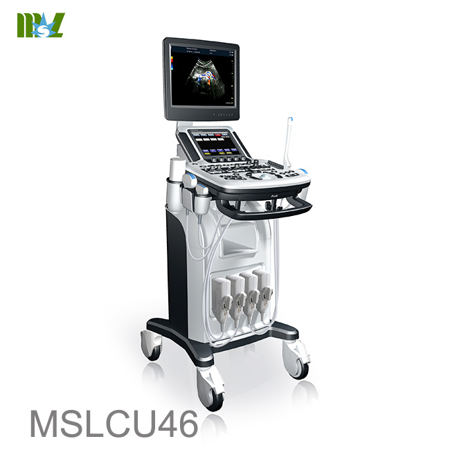
Hot sale Sonoscape ultrasound | Chison ultrasound price list
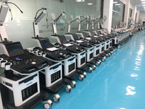
MSL TEAM picture


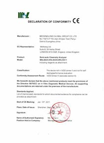
MSL Certificate
MSL Medical cooperate with DHL,FEDEX,UPS,EMS,TNT,etc.International shipping company,make your goods arrive destination safely and quickly.


 Price is 8-20% Lower Than Other