|
Serial number
|
Main Technical Specifications
|
|
1
|
Flat Panel Detector
|
|
1.1
|
Amorphous silicon flat panel detector
|
|
1.2
|
Effective pixels:≥9 million
|
|
1.3
|
Pixel matrix:≥3070*3070 pixels
|
|
1.4
|
Quantization depth:≥16bit
|
|
1.5
|
Effective area:17”*17”
|
|
1.6
|
Spatial resolution:≥3.7Lp/mm
|
|
1.7
|
Imaging time:≤7s
|
|
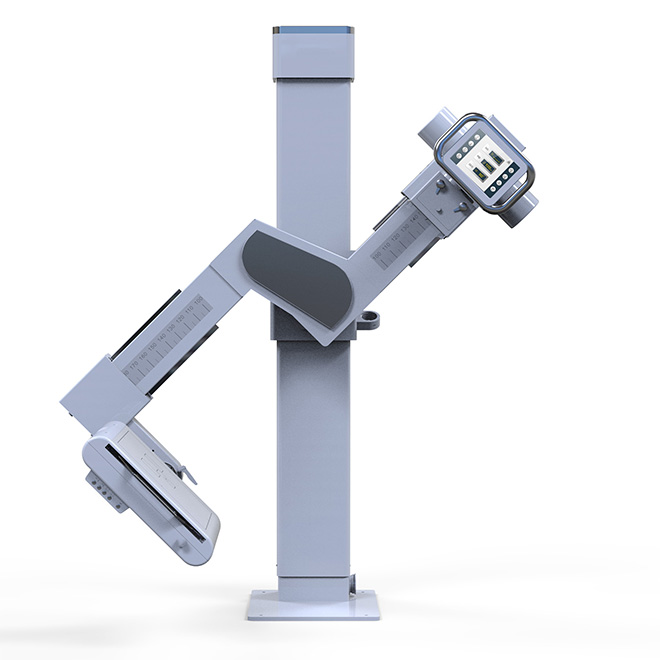
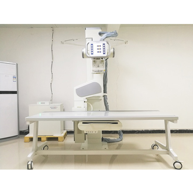
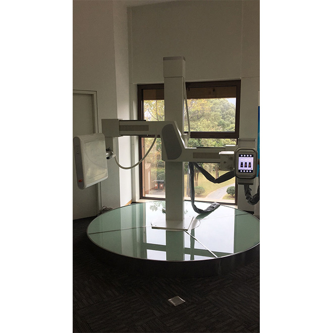
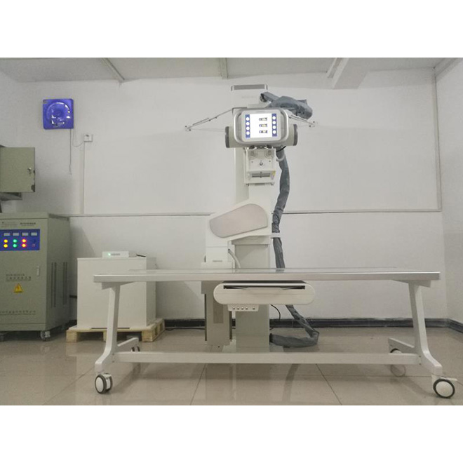
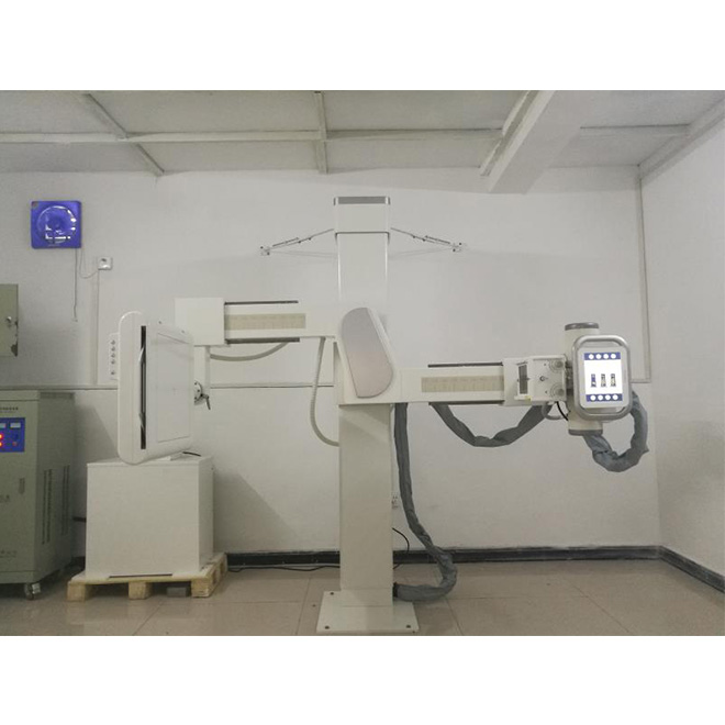
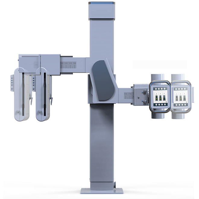
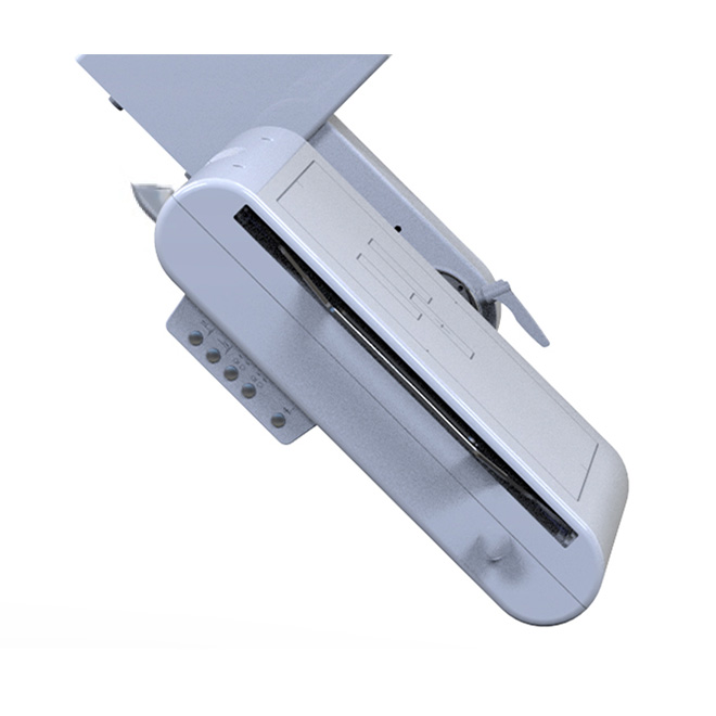
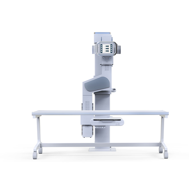
2
|
Mechanical structure: scythe arm(type Z)
|
|
2.1
|
Vertical range(scythe arm):600mm ~ 1600mm(±5%)
|
|
2.2
|
Rotation range of the scythe arm:-45°~ +135°(±2°)
|
|
2.3
|
SID (vertical and horizontal)can be adjusted electronically,the range:1000mm~1800mm(±5%)
|
|
2.4
|
Rotation range of the tube assembly :-15°~ +15°(±2°)
|
|
2.5
|
Rotation range of the detector :-45°~ +45°(±2°)
|
|
2.6
|
The detector end can independently control the movement of the stand
|
|
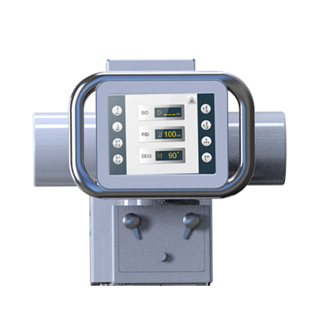
2.7
|
9.5 "LCD touch screen
|
|
2.8
|
Minimum height of the equipment room:2.4m
|
|
2.9
|
The tube and detector move synchronously to maintain the balance of the stand
|
|
3
|
High Voltage Generator
|
|
3.1
|
High voltage generator smart network voltage real-time monitoring system(smart monitor)
|
|
3.2
|
Power:≥50kW
|
|
3.3
|
kV range:40kV-150kV
|
|
3.4
|
mA range:10mA-630mA
|
|
3.5
|
mAs range:0.1mAs-630mAs
|
|
3.6
|
Maximum operating frequency:200kHz
|
|
4
|
X-ray Tube
|
|
4.1
|
Focus size:≤0.6mm/1.2mm
|
|
4.2
|
Focus power:
small focus:≥ 20kW
large focus:≥ 50kW
|
|
4.3
|
Maximum heat capacity:≥300kHU
|
|
4.4
|
Maximum peak voltage:≥150kV
|
|
5
|
Filter grid
|
|
5.1
|
Ratio:≥10:1
|
|
5.2
|
Density:≥40L/CM
|
|
6
|
LED Collimator
|
|
6.1
|
Time limit of indicator :30±3s
|
|
6.2
|
Inherent filtration:1.0 mm Al.EQ.
|
|
7
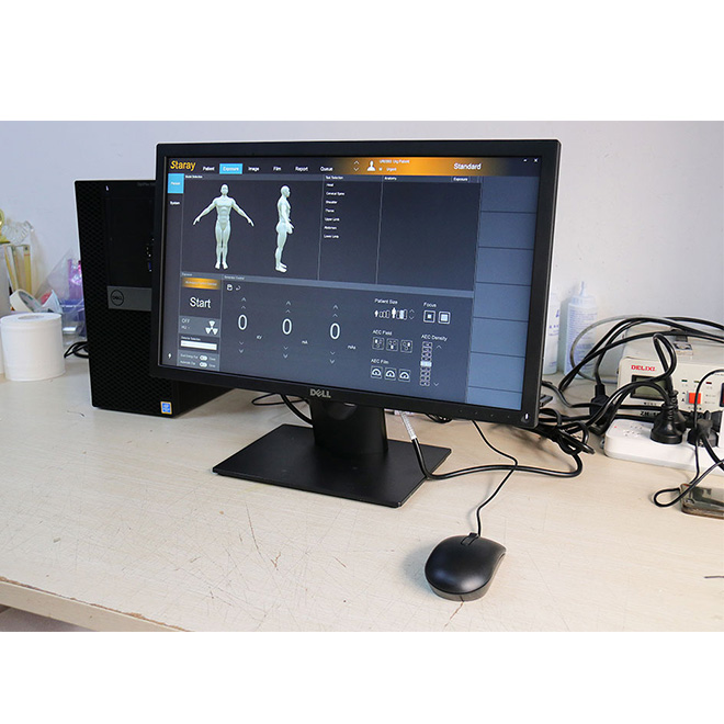
|
Workstation
|
|
7.1
|
DELL 5040MT,Intel Pentium G4400 processor,
4G/500G/DVDRW/WIN10,E2216H 21.5 inch high contrast display
|
|
8
|
Software function(patient management)
|
|
8.1
|
Remote direct access patient information in RIS with worklist protocol;
Manually create the patient examination information;
Emergency quick registration;
Simple,advanced, or custom data query methods;
The image CD burning in Dicom standard ,can be viewed on any standard image workstation;
Inquiry and management of historical image data;
Detection disk space, automatic cleaning of old check data;
Dicom transmission of image and seamless connection with PACS;
Support diagram text report,carry report library;
Print patient's reports, exposure times can be displayed in the list of patients;
The original image is easy to view.
|
|
9
|
Software function(software direct exposure control)
|
|
9.1
|
Support customization for dose template;
Selection part and position template, automatically bring out the matching dose; Selection shape of the patient, automatically select the matching dose, and adjust to the dose online;
Control the size of focus;
Display generator's status transparent;
Refusal and acceptance of images;
Support automatic sending of images.
|
|
10
|
Software function(image processing)
|
|
10.1
|
Supports dual or multi-screen images display;
Manually/automatically/preset window width and window level, partial window width and window level;
Operation positive and negative of image,flip image,rotation image,scaling image and roaming;
Automatic or manual addition of image information;
Line, angle, rectangle, ellipse, polygon and other measuring tools;
Balance tissue,enhancement contrast,optimization dose;
Enhancement edge : automatic recognition and analysis of images,enhance edge sharpness.
|
|
10.2
|
Preview the post-processing image 1:1: the doctors make more intuitive diagnosis in image optimization;
CAD View: just click the mouse ,doctors can see the details
|


 Price is 8-20% Lower Than Other