


 Price is 8-20% Lower Than Other


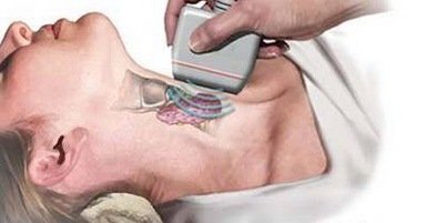
Thyroid B-mode ultrasonography is a method for examining thyroid, parathyroid glands, and other related lesions.
Normal value
(1) Thyroid thickness (anteroposterior diameter) and width (right and left diameter) Standard measurement section In the series of thyroid cross sections, the thickest and widest thyroid parenchyma is selected as the standard section, and the probe pressure should be as light as possible. Measurements were taken at the edge of the hyperechoic line at the thickest and widest thyroid of the thyroid.
The normal adult reference value (cm) is about 1.5-2.0 cm in the two leaves and 2.0-2.5 cm in width, and the isthmus thickness is less than 0.5 cm. (2) Longitudinal diameter (upper and lower diameter) measurement standard of thyroid. Measurement section In the series of longitudinal sections of thyroid, the longest part of thyroid parenchyma is selected as the standard section, which requires the probe pressure to be as light as possible. The measurement position was selected on the edge of the hyperechoic line at the longest position of the thyroid gland. Normal Adult Reference (cm): Left and right leaf lengths are 4.0-6.0 cm, and isthmus length is 1.5-2.0 cm.
Clinical significance
Abnormal results: (1) determine whether the mass is located in the thyroid gland, which is diffuse or localized; (2) identify the mass is cystic or solid; (3) determine the mass is single or multiple; (4) It can be judged whether the tumor is benign or malignant; (5) The efficacy can be followed up after surgery or after the use of the drug; for nodules that cannot be seen, ultrasound can find the number of nodules and nodules >=0.5 cm. The sensitivity of ultrasound diagnosis of primary hyperparathyroidism was 66%-88.4%. Color Doppler ultrasound can not only improve the sensitivity of ordinary two-dimensional ultrasound on the detection of small parathyroid lesions, but also help to locate the diagnosis, and easy to identify with the thyroid and lymph nodes. The
Subjects to be examined: (1) goiter; (2) thyroiditis; (3) thyroid tumor; (4) thyroid cyst; (5) cervical lymphadenopathy; (6) parathyroid adenoma and parathyroid hyperplasia (7) Multiple endocrine tumors; (8) Parathyroid carcinomas.


